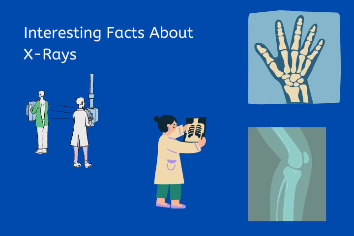
We all know X-rays are a type of energy that can pass through our bodies. They are often used by doctors to take pictures of our bones or to look for something foreign in our bodies. But there is a lot more to X-rays than just that! Here are 20 interesting facts about X-rays that you probably didn’t know.
- The first X-ray was taken by Wilhelm Conrad Roentgen in 1895. He discovered them completely by accident while experimenting with electricity in his laboratory.
- X-rays are a form of electromagnetic radiation, like visible light and radio waves. They have a shorter wavelength than visible light and can penetrate human tissue. X-rays are used in medicine to create images of the inside of the body. They are also used in airport security scanners and to detect defects in metal objects.
- Roentgen named them “X-rays” because he wasn’t sure what they were. The “X” stands for the unknown.
- Knowing how much energy is needed to produce an X-ray can help scientists understand the temperatures and densities of the stars and other objects in space.
- Some materials will absorb more X-rays than others. This is why lead aprons are often worn when having an X-ray taken, to protect us from too much exposure to the radiation.
- Bone and teeth are denser than soft tissues like skin and muscles, so they appear white on an X-ray image. Fatty tissues appear gray and air appears black.
- Metal objects will also show up very clearly on an X-ray image, which is why they are often used to find coins and other metal objects that have been swallowed by people or animals.
- Although we usually think of them as being invisible, we actually emit low levels of X-rays all the time just from being alive! Unlike visible light, which our eyes can see, we cannot seeX-rays with our eyes because they have too much energy for us to detect them directly.
- When Wilhelm Roentgen announced his discovery of x-rays in 1895, it didn’t take long for European surgeons to start using the new technology to find foreign objects in a human’s body. A British doctor was one of the first to make a diagnosis and found a needle embedded in a woman’s hand.
- In the early days of x-ray technology, people were unaware of the dangers posed by this new form of radiation. It was thought that x-rays were harmless, and people routinely exposed themselves to high levels of radiation without any ill effects. However, we now know that x-rays can cause serious damage to the body, and prolonged exposure can lead to cancer. Today, we take precautions to limit our exposure to x-rays, and we are much more aware of the risks posed by this powerful form of radiation.
- Yes, it’s true: the very same thing that can cause cancer can also cure it. Ultraviolet rays, whether from the sun or a tanning bed, can cause skin cancer. But doctors have also been using UV light to treat cancer since the early days of x-rays. In fact, even back in 1895, when Wilhelm Roentgen discovered x-rays, doctors were already using them to burn off moles. So while overexposure to UV rays can be dangerous, there’s no denying that they can also be therapeutic.
- Did you know that diamonds don’t show up on X-rays? This is because they’re made of carbon, which has a very low atomic number. This means that the atoms in diamonds don’t interact with X-rays very much, so they appear nearly transparent.
- Electromagnetic radiation travels at the speed of light in a vacuum. This includes x-rays and radio waves. In a vacuum, there is no medium for electromagnetic radiation to travel through, so it travels at its fastest possible speed.
- In 2009, the Science Museum of London held a poll to determine the most important modern scientific discovery. The winner, by a wide margin, was the X-ray. Even penicillin, which has saved countless lives, came in second.
- There are two types of x-rays: hard x-rays and soft x-rays. Hard x-rays have higher energy than soft x-rays. Hard x-rays are used to image dense tissues, like bone. They can penetrate through the body and cast a shadow of the structures inside onto film. Soft x-rays have lower energy than hard x-rays. Soft x-rays are used to image soft tissues, like organs. They can also be used to take pictures of the inside of the body, but they don’t penetrate as far as hard x-rays. Soft x-rays usually don’t need to be filtered as much as hard x- rays, so they don’t produce as much radiation exposure.
- Thomas Edison is perhaps best known for his work with electricity, but he also made significant contributions to the field of X-ray research. However, after sustaining damage to his eyesight from exposure to X-rays, Edison abandoned his work in this area. His assistant, Clarence Dally, continued Edison’s research, but he ultimately paid a great price for his dedication. After years of working with X-rays, Dally developed cancer in both arms and eventually had them both amputated.
- In the 1980s, there was an X-Ray machine that would randomly give patients overdoses of radiation due to a programming error. This resulted in many deaths, and the hospital had to switch to a new machine.
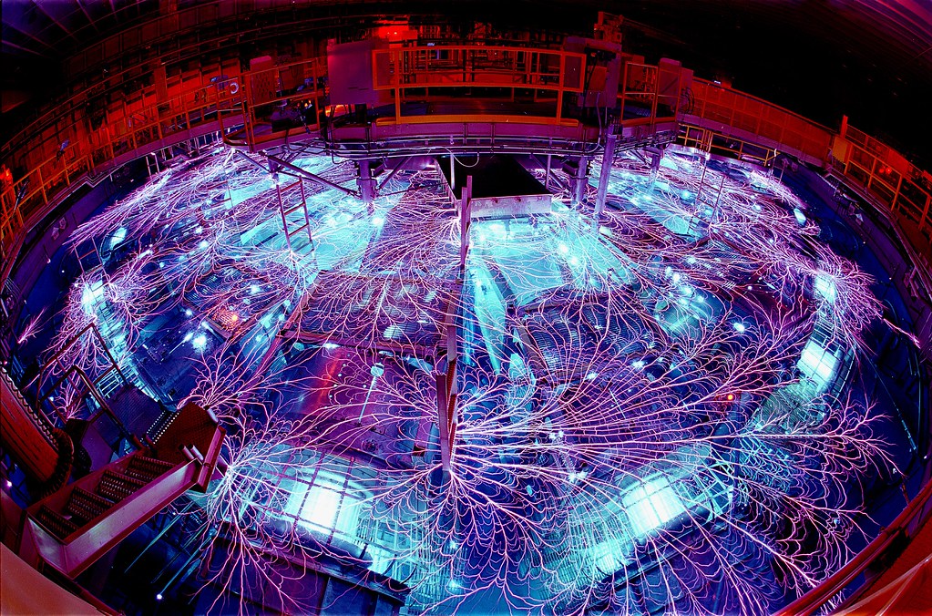
- You might not expect to find the world’s most powerful X-Ray generator in the middle of the desert, but that’s exactly where you’ll find the Z machine. Located in New Mexico, this massive machine is capable of producing temperatures greater than 6 billion degrees Fahrenheit. The Z machine is used to study the behavior of matter under extreme conditions, and it has contributed to our understanding of astrophysical phenomena such as supernovas. It’s also been used to create new materials with unique properties.
- It’s no secret that Vincent Van Gogh was a bit of an eccentric. Even those who are familiar with Van Gogh’s idiosyncrasies may not know that he was also prone to painting over his own work. Many of his most famous paintings were hidden for years, covered up by later works. It wasn’t until the development of X-ray technology that art experts were able to see the hidden beauty beneath the surface.
- A CT scan is a type of imaging test that uses X-rays and a computer to create detailed images of the inside of your body. A chest CT scan specifically creates images of your lungs, heart, and blood vessels. While a chest X-ray also uses X-rays to create images of your chest, a CT scan is much more detailed. As a result, compared to chest X-rays, a chest CT scan exposes patients to 250-350% more radiation.
X-rays are a fascinating type of energy that have many uses both inside and outside of the human body! Now that you know a little bit more about them, keep your eyes peeled for opportunities to learn even more about these amazing phenomena!